Technical Note 182

DeNovix Acridine Orange Assay Protocol

Introduction

Acridine Orange (AO) is a fluorescent dye that enters live and dead cells and interacts with DNA and RNA by intercalation or electrostatic attractions. When bound to DNA, it has an excitation maximum at 502 nm and an emission maximum at 525 nm.

Although AO enters dead cells, the intensity of the green fluorescence is very low compared to the green fluorescence emitted from live cells. This app may be useful when counting solutions containing either red blood cells and/or significant cellular debris. The DeNovix AO Assay and AO app on CellDrop™ Series instruments enable rapid automated cell counting for cell suspensions.

Kit Contents

Kits include a solution of AO in PBS. The AO reagent may be stored protected from light at 2 – 8oC in an airtight container.

Assay SizeNumber of Tests
0.25 mL50
1.5 mL300

Sample Prep

  1. Equilibrate all solutions to room temperature, and vortex before use.
  2. For each sample, mix AO and a cell suspension together in a 50% solution.
    • Note: A 50% solution is made up of one part AO and one part cell suspension, resulting in a Dilution Factor of 2 for the cell suspension.
  3. Mix sample thoroughly prior to loading onto the CellDrop.

Sample Measurement

  1. Ensure that the arm is in the down position and launch the AO app.
  2. Clean the sample surfaces if there is visible debris in the preview image.
  3. Optional: Enter a sample name and additional sample information.
  4. Select or create a protocol, and enter the Dilution Factor of 2 for a 1-to-1 dye/cell suspension mix.
  5. With the arm down, dispense the sample aliquot into the measurement chamber using the groove on the lower sample surface as an alignment guide.
    • Note: The volume of sample required depends on the protocol settings for the chamber height. The required volume is displayed on the Count button.
  6. Adjust the focus and exposure. Verify focus in green channel prior count.
  7. When the cells have settled and are no longer moving, tap the Count button.
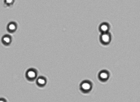
Figure 1: Correct focus will show live cells with bright white centers and sharp black rings.
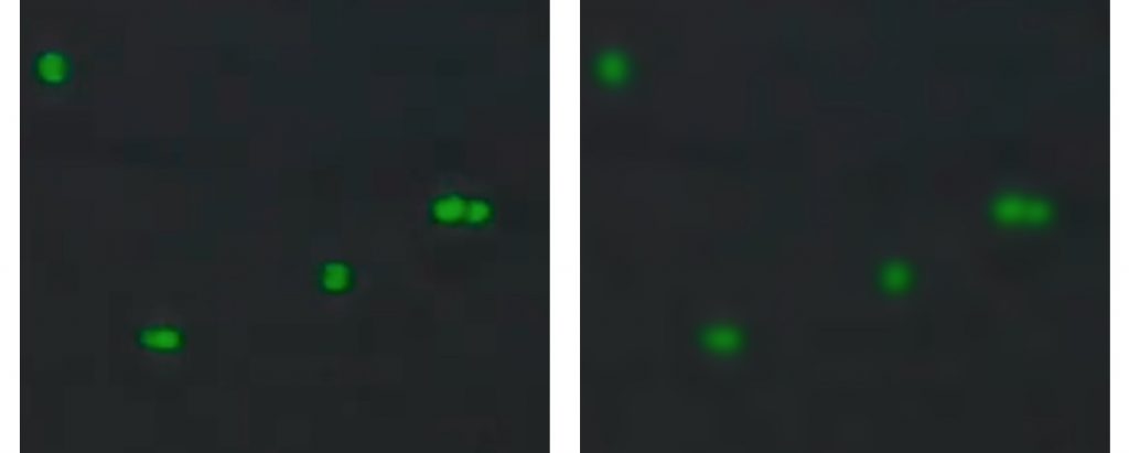
Verify Green Channel Focus. Left - good focus, Right - out of focus.
Figure 1: Verify Green Channel Focus. Left - good focus, Right - out of focus.

Best Practices

  • Ensure that both upper and lower sample surfaces are clean.
  • Lower the arm prior to dispensing a sample onto the measurement surface.
  • Avoid introducing air bubbles.
  • Use the correct dilution factor in the protocol settings.
  • Adjust the focus in the brightfield channel so that cells have bright white centers and a sharp black ring as seen in Figure 1. Verify focus using the green channel checking that cells have a sharp outline (Figure 2.)
  • Adjust exposure in the green channel so that fluorescent cells are not over or underexposed, as shown in the info dialog in the exposure menu.

Refer to Technical Note 186 – CellDrop Best Practices for additional guidance.

Refer to denovix.com/sds for safety data sheets for CellDrop Cell Counting Assays.

01-MAR-2023