Based in the Department of Biology at the University of Utah, the Jorgensen laboratory is home to a uniquely decorated DeNovix DS-11 FX+ Spectrophotometer / Fluorometer (see image below!).
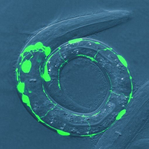
The Jorgensen laboratory, established in 1994 by Dr. Erik Jorgensen, studies the molecular basis of synaptic transmission, with a strong record of developing new methods to address long standing questions. They have developed methods to generate both random and targeted insertions in their main model system, the nematode C. elegans. They pioneered superresolution microscopy to localize and track single proteins. Further, they developed “zap-and-freeze” electron microscopy to take nanometer-scale pictures of neuronal synapses within 5 milliseconds of a transmission event. The eventual goal will be to fuse these methods to characterize the molecular topography of the cell in time and space.
In a paper recently published in eLife, Brian Mueller et al. applied all of these new methods to describe how two different calcium channels collaborate at synapses to generate robust and tunable transmission from nerves to muscles.
The team used the DeNovix DS-11 to quantitate DNA for routine cloning as well as for creating transgenic worms.