In parallel with advancements in next-generation sequencing, the number of published studies using isolated nuclei has risen steeply recently. The development of more affordable nuclei isolation and sequencing techniques enables us to decode the gene regulatory mechanisms that characterize cell fates. Single- nuclei sequencing reveals the unique and heterogeneous characteristics of different cell populations, having many biological, biomedical and clinical applications.1
Brain Tissue Isolated Nuclei
Particular attention has been given to disentangling the genetic mechanisms behind brain cell architectures and functions. As nuclear transcriptomes mirror cell cytosolic profiles, nuclei isolation from brain tissue samples is a valuable tool for characterizing the genetic makeup of different brain cell types, identifying where it differs and where it is conserved between cell populations.
Understanding differences between transcriptomes in different physiological states using isolated nuclei DNA is vital for understanding complex diseases underpinned by multiple genes.2 An advantage of single-nuclear sequencing is that isolated nuclei are more stress resilient compared to cells, and the implementation of brain tissue nuclei isolation has proven invaluable for human brain tissue analysis.3
Choosing the correct protocol for nuclei isolation is critically important for maintaining nuclear RNA and protein integrity, which is fundamental in downstream processes like RNA sequencing and proteomics. Quantification of the isolated nuclei is an essential step in the protocol. See how DeNovix and 10x Genomics methods complement one another to provide powerful, yet affordable, methods for quantifying isolated nuclei concentration from brain tissue.
Quantifying Isolated Nuclei Concentration
A recent study designed a protocol for the isolation of genetic nuclei from the brain tissue of adult African turquoise killifish to study the molecular mechanisms behind brain aging. This step-by-step tailored protocol was adapted from a 10x Genomics protocol for mice brain-isolated nuclei and highlights the use of DeNovix in producing isolated nuclei adequate for downstream single-cell experiments.4
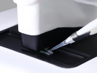
DeNovix CellDrop Automated Cell Counters™
After using a Dounce homogenizer to mechanically dissociate the nuclei, followed by a series of centrifugation steps to purify the nuclei, isolated nuclei DNA concentrations were then quantified using the DeNovix CellDrop™. Samples were resuspended in nuclei wash buffer solution and small samples of this suspension were examined by the DeNovix cell counter. This step is vital in counting the number of isolated nuclei, and for determining the relative proportion of multiple nuclei, to ensure the correct nuclei density for the next steps in the protocol.4
Nucleic acid staining with propidium iodide enables singlet nuclei to be differentiated from multiple nuclei and debris. The authors provide detailed instructions on the types and quantities of reagents, along with any necessary materials, required to accurately evaluate DNA concentration in killifish brain isolated nuclei.4
10x Genomics Chromium Controller
Once the DNA concentration was accurately assessed the authors then used the 10x Genomics Chromium Controller to process the isolated nuclei for single-nucleus RNA sequencing. This technique produces single-cell gel beads in emulsions, termed GEMs, that provide a solid support for isolated nuclei. The resulting 10x Genomics Chromium Controller GEMs contain the isolated nuclei and all the reagents needed for reverse transcription.4
Together, the DeNovix and 10x Genomics protocols allowed for successful extraction of isolated nuclei for single-nucleus RNA sequencing. The concentration quantified using the DeNovix cell counter determines the sample input for the 10x Genomics Chromium Controller, informing how samples are processed in the 10x Genomics device. Thus, these techniques worked hand-in-hand to form the first protocol for high-quality isolated nuclei from African turquoise killifish brain tissue.4
Isolation of the killifish brain nuclei in this study displays the crucial roles and complementary utility of DeNovix’s and 10x Genomics’ tools in nuclei isolation. This empirically optimized protocol was the first to isolate high-quality nuclei from killifish brain tissue, providing a vital step towards the generation of high-quality single nuclei omic libraries for model vertebrates. Although tailored to this organism the authors note that this protocol can be utilized for other tissue types and other organisms, enhancing the application of these methods to other tissues and systems.
If you would like to find out more about the DeNovix CellDrop visit the product page. You may also be interested in our nuclei protocol settings or our tech note on counting isolated nuclei using the CellDrop.
References
- Butto, T., Mungikar, K., Baumann, P., Winter, J., Lutz, B. and Gerber, S. 2023. Nuclei on the Rise: When Nuclei-Based Methods Meet Next-Generation Sequencing. Cells. 12(7), p.1051.
- Maitra, M., Nagy, C., Chawla, A., Wang, Y.C., Nascimento, C., Suderman, M., Theroux, J.F., Mechawar, N., Ragoussis, J. and Turecki, G. 2021. Extraction of nuclei from archived postmortem tissues for single-nucleus sequencing applications. Nature Protocols. 16(6), pp.2788-2801.
- Kulkarni, A., Anderson, A.G., Merullo, D.P. and Konopka, G. 2019. Beyond bulk: a review of single cell transcriptomics methodologies and applications. Current opinion in biotechnology. 58, pp.129-136.
- Teefy, B.B., Adler, A., Bhala, R. and Benayoun, B.A. 2022. Gentle Isolation of Nuclei from the Brain Tissue of Adult African Turquoise Killifish, a Naturally Short-Lived Model for Aging Research. JoVE (Journal of Visualized Experiments). 186, p.e64165.